Study reveals key immune cells linked to relapse in NMO
A new study has revealed how specific immune cells in the cerebrospinal fluid (CSF) of patients with neuromyelitis optica (NMO) may drive disease activity, offering new insight into the mechanisms behind this serious autoimmune condition. The findings suggest that a distinct population of B cells in the central nervous system may be responsible for producing harmful antibodies, and could serve as both biomarkers and therapeutic targets.
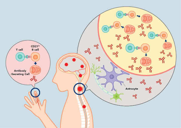
NMO is a rare but debilitating autoimmune disease marked by relapsing inflammation of the optic nerves and spinal cord. This inflammation is triggered by antibodies that attack aquaporin-4, a protein found on the surface of astrocytes in the central nervous system. As lead research Dr. Ryusei Nishigori explains, “While highly effective therapies have been developed to reduce relapse rates, the treatments are often expensive and can increase the risk of infections, posing challenges for long-term management.”
To address this, the research team sought to understand where and how these pathogenic antibodies and the immune cells that produce them are generated. They collected and compared immune cell profiles from both blood and CSF in patients with NMO, multiple sclerosis (MS), and other neurological diseases, using high-parameter flow cytometry. This method allows various immune cell markers to be measured, both on the surface and inside the cells, giving a detailed overview of immune activity in different areas of the body.
“It was unclear how and where harmful antibodies and immune cells are produced in NMO,” Dr. Nishigori noted. “This lack of understanding hindered the development of more refined targeted therapies and personalized treatment strategies. We addressed this by analyzing immune cells in paired blood and CSF using high-parameter flow cytometry.”
Their analyses showed that in NMO patients, particularly during acute attacks, CD21lo B cells accumulate in the CSF at significantly higher levels than in the blood. These B cells are activated in response to an immune trigger (such as a pathogen or, in the case of autoimmune diseases, the body’s own tissues) and can produce antibodies. In the study, the CSF CD21lo B cells exhibited surface markers associated with tissue residency and inflammatory function, including CD69 and CXCR3, as well as high expression of the C5a receptor.
Functional experiments confirmed that these cells can respond to C5a, a complement component known to be elevated in NMO. C5a signaling promoted their differentiation into antibody-secreting cells (ASCs), which in turn produced aquaporin-4-specific antibodies. This finding not only connects B cell activity in the CSF to the core mechanism of NMO pathology but also implicates C5a signaling as a potential therapeutic target.
The researchers also observed a correlation between the frequency of CD21lo B cells in the CSF and patients’ annual relapse rates, suggesting that these cells may serve as biomarkers of disease activity. Furthermore, these patterns were not observed in MS or other neurological diseases, highlighting the specificity of this immune signature for NMO.
In future work, the team plans to assess the clinical utility of CD21lo B cells as biomarkers for disease activity and relapse prediction. They also aim to conduct longitudinal studies in patients who do not respond to current treatments, tracking immune changes in the CSF over time to identify indicators of treatment response and disease progression.
The study offers a new framework for understanding the immunology of NMO, highlighting the importance of analyzing CSF, not just blood, when studying central nervous system autoimmune diseases. “Understanding the immune cells responsible for disease may lead to safer and more effective treatments,” Dr. Nishigori adds. “We hope this research will help improve the lives of people with NMO.”
書誌情報
Nishigori, R., Hamatani, M., Yoshitomi, H., Kimura, K., Takata, M., Ashida, S., Fujii, C., Ochi, H., Takahashi, R., Kondo, T., & Ueno, H. (2025). CD21lo B cell subsets are recruited to the central nervous system in acute neuromyelitis optica. Brain. [DOI: 10.1093/brain/awaf086]